
3D Imaging
Advanced Oral Surgery Diagnostics In Caldwell & Florham Park NJ
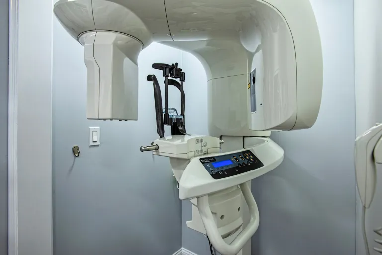
Carestream 9300 Cone Beam CAT Scan
The NJ Center for Oral Surgery is proud to offer the Carestream 9300 Select 3D Cone Beam CAT Scan.
CAT images provide Dr. Jacobs, Dr. Kirsch and Dr. Levin with the most comprehensive and accurate information available to determine bone quantity and quality. The dental scan will provide the exact location of anatomical structures, the contours of the jaw bone, and the best sites for your implants. With Dental CAT Scanning implant procedures can be planned in detail before any surgery takes place. This means virtually no surprises when a doctor performs surgery.

Key Benefits of Cone Beam CAT Scan
- Our Cone Beam CAT Scan emits much less radiation compared to a regular CAT Scan found at hospitals and radiology facilities. In some cases it can be 10x less.
- Allows our surgeons to visualize internal anatomy that can not be diagnosed externally or by clinical examination.
- Plan treatment and surgery by providing a 3D detailed map to the area of interest.
- Assess risks for a surgery by providing detailed anatomy and visualization of vital nerves and other structures. This may be particularly important when evaluation impacted teeth and sites for dental implants.
- Analyze the position and orientation of critical structures, like nerves, teeth roots, implant sites, and the sinus.
- Our CAT Scan provides 3D images vs. traditional x-rays which only provide 2D.
- Unlike regular x-rays, our CAT Scan can see the differences between many types of tissues including bone, teeth, nerves and soft tissues.
- CAT Scans are noninvasive, and can eliminate the need for exploratory surgery in many cases.
- CAT Scans can identify the size, extent and effects of conditions such as infections, cysts and tumors.
- Much faster scan time 10-40 seconds, while a regular CAT Scanner takes several minutes to complete.
- Our fee can be 50% less expensive than a CAT Scan obtained at a hospital or radiology facility.
- “If the wisdom tooth is deemed to be in close proximity to the mandibular canal, then a CBCT or CAT Scan should be ordered for further evaluation. A single panoramic radiograph could be insufficient owing to image disturbance compare with a CAT Scan. However, CBCT is the gold standard owing to lower radiation exposure and lower cost compared with CT.” J Oral Maxillofacial Surgery 73:1672-1685, 2015
3D Imaging FAQ
The Evaluation Stage

What to Expect During Consultation
During your initial visit, our oral surgeon will thoroughly review your medical history and discuss any specific concerns you have about your oral health. We’ll perform a comprehensive examination and explain why 3D imaging is recommended for your particular case. You’ll learn how this advanced technology helps us provide safer, more precise surgical care.
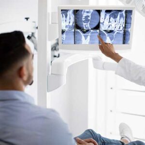
We’ll walk you through sample 3D images similar to what we’ll capture of your oral structures, helping you understand exactly what we’re looking for and how the scans influence your surgical plan. Our team will also discuss timing, costs, and insurance coverage for both the imaging and your planned procedure.
Exam Process
What to Expect During the Imaging Procedure
The 3D imaging process is quick, comfortable, and completely painless. You’ll sit in an open scanning chair while our imaging system rotates once around your head, capturing hundreds of detailed images in just 14 seconds. Our experienced technologist will ensure you’re properly positioned and comfortable throughout the brief scan.
There’s no special preparation needed, and you can breathe normally during the scan. Unlike traditional medical CT scanners, our dental imaging system is open and non-confining. The entire process, including positioning and image capture, typically takes less than 5 minutes to complete.
Post-Exam Information
What Happens After 3D Imaging?
There is no recovery period required after 3D dental imaging – you can immediately resume all normal activities, including eating, drinking, and returning to work or school. Our oral surgeon will analyze your scans immediately, often discussing the findings with you during the same visit. We’ll use these detailed images to show you exactly what we’ve found and explain our recommended surgical approach.
Using your 3D scans, we’ll create a comprehensive surgical plan tailored to your specific anatomy and needs. The detailed images become part of your permanent medical record, allowing us to track changes over time and share important information with your other healthcare providers when needed.
We’ll provide you with a clear treatment timeline and detailed pre-surgical instructions based on your specific procedure. Our surgical coordinator will help you schedule your procedure and answer any questions about insurance coverage or financing options. Throughout the entire process, we remain committed to ensuring you feel informed, comfortable, and confident about your upcoming oral surgery.